Engineering tissues for transplantation and disease modelling
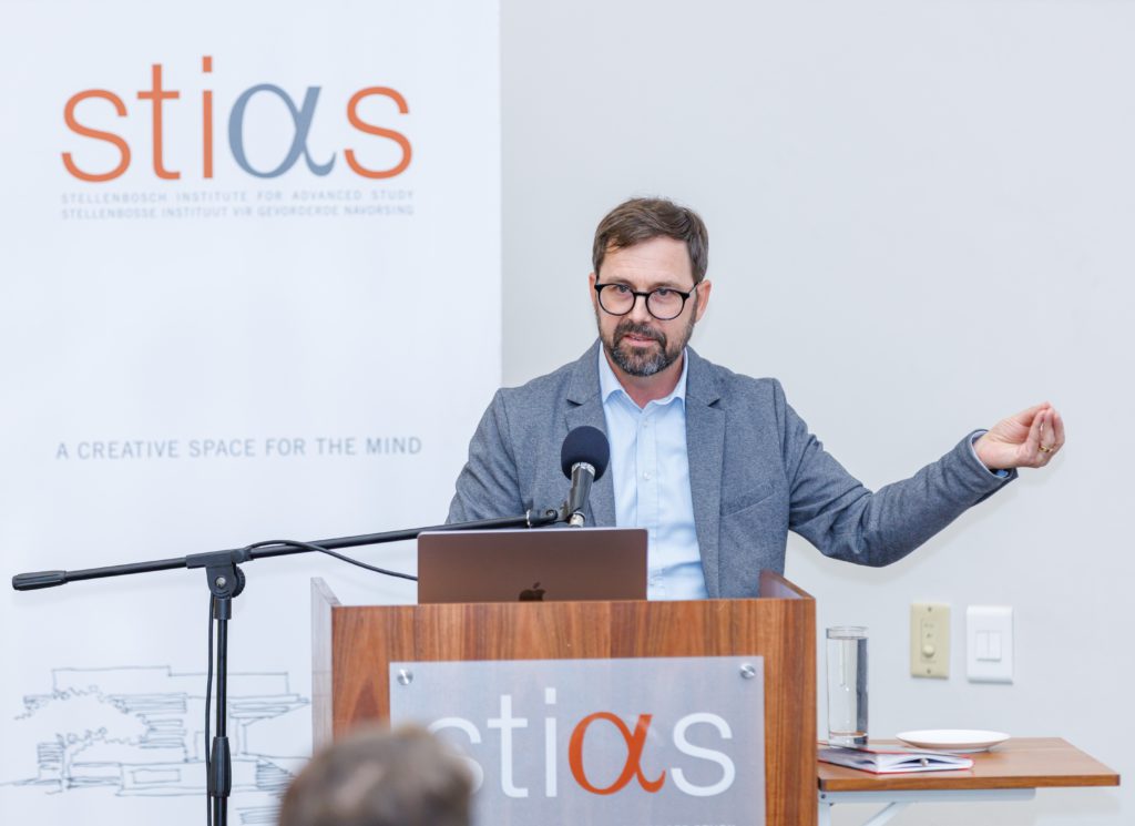
“By engineering human tissues in the lab, we aim to accelerate drug discovery, develop personalised treatments, and pave the way for future regenerative therapies. Some of this work is in preclinical trials – I believe will see interesting progress quite soon,” said Daniel Aili in the third STIAS public lecture of 2025.
Daniel Aili is Professor of Molecular Physics in the Division of Biophysics and Bioengineering at Linköping University (LiU), Sweden. After postdoctoral research at Imperial College London and Nanyang Technological University in Singapore, he was recruited to LiU, where he established the Laboratory of Molecular Materials. His interdisciplinary research focuses on bioresponsive materials for biomedicine and bioengineering. He has published about 100 peer-reviewed papers, authored five book chapters, and holds over eight patents.
Aili is also an active entrepreneur and scientific advisor for several start-ups. He has been awarded the AkzoNobel Nordic Prize for Surface and Colloid Chemistry, the Arnbergska Prize from the Royal Swedish Academy of Sciences, a Future Research Leader Award from the Swedish Foundation for Strategic Research, and is an elected member of the Swedish National Committee for Molecular Biosciences. He is a Knut and Alice Wallenberg Academy Fellow and holder of an ERC Consolidator Grant. At LiU, he leads a research team of approximately 20 students and senior researchers.

Not axolotls
Aili explained that unlike axolotls (a fresh-water salamander) humans cannot regenerate lost limbs or organs. “Axolotls have an amazing capacity that humans lack – they can heal by regeneration. If they lose an organ or limb, it will regrow and look the same as the original. Humans can’t do this.”
“Some human tissue can regrow − like liver − but not our limbs,” he continued. “Human tissues heal by scarring which is evolutionary – to heal quickly and restore barrier function to prevent infection. But scar tissue is not the same as normal tissue – it has a different architecture and function. If we have an injury to the heart in a heart attack the scar tissue impairs the contraction so there is a loss of function. It’s similar in the central nervous system.”
He pointed out that all humans originate from a single cell that, during embryonic development, differentiates and multiplies into more than 30 trillion cells – “100 times more than the stars”. They specialise into over 200 different types (including epithelial, connective tissue, muscle, nervous system, blood and immune, reproductive, stem and progenitor cells) which spontaneously organise into functional tissues and organs of astonishing complexity.
Although all cells in the human body carry the same genetic information as the first few cells in the embryo, our ability to regenerate lost or damaged tissue is severely limited. “Why can’t we regenerate? We don’t have the answer as yet,” said Aili. “The blueprint is there – we have DNA that codes for the whole body, the same DNA as in the first cell. But as the embryo develops the regeneration function is lost.”
“However, by combining cells, biological factors, and supporting biomaterials, researchers can now grow structures that resemble tissues and organs in the lab,” he continued. “These lab-grown tissues have already become invaluable tools for biomedical research and drug development and hold great promise for regenerative medicine.”
Closer to human models
The challenge lies in creating models in the laboratory that can mimic all the complexities of the human body. Aili discussed his research group’s efforts to design biomaterials and biofabrication technologies for creating novel regenerative strategies and physiologically relevant disease models. “We develop soft, highly hydrated materials that not only provide structural support but also deliver biochemical and mechanical signals to guide cellular organisation. Using advanced 3D bioprinting techniques, we can create well-defined and more complex, tissue-like architectures.”
To do this they are engineering materials that mimic the extracellular matrix (ECM). The ECM is like a cellular soup. It’s the biological material that cells reside in and consists of about 200 or so different molecules including core structural proteins, adhesive glycoproteins, growth factors and signalling molecules, remodelling enzymes and inhibitors. It keeps cells in their 3D structure but also facilitates cell adhesion, cell-to-cell communication and differentiation.
“ECM is hard to handle and process and exceptionally hard to replicate,” said Aili. To make synthetic ECM they are using a reductionist approach – starting with one component and adding more. “In this way we can build a library of the different designed components that, when combined, mimic various aspect of the ECM.”
Getting the combination of cells and materials right enables them to start to mimic the spatial complexity of human tissues and provide the right environmental conditions so that when stem cells are added they can develop into something that looks and behaves like tissues and even organs. These engineered materials to culture cells and tissues have been used to model cardiac and liver organoids, neural 3D models, and 3D breast-cancer models.
Aili noted that currently, according to the United States’ National Institutes of Health, 90% of new drugs validated by animal testing fail in clinical trials for use in humans. Physiologically relevant human disease models can therefore greatly facilitate drug development.
“We are not rats and don’t have the same responses,” he said.
Breast-cancer models
Aili’s described some of his group’s work in developing 3D models for breast cancer.
The failure rate for cancer drugs is particularly high with about 97% failing in clinical trials. “This is very expensive but obviously also a major problem for the patients urgently needing therapies.”
Basically, the current standard methods when testing drug safety and efficacy is to use human cells cultured as thin layers on plastics – which is a different environment to tissues, or to test them on animals. In addition to ethical problems, animals can respond very differently to the drugs.
“We are therefore trying to make better disease models that behave more like 3D human cancer tissues. But tumours are not just cancer cells – they contain immune cells, blood vessels, pericytes and stromal cells which all contribute to disease progression. We are trying to find ways to replicate this complexity.”
They have therefore used biofabrication strategies, 3D printing and their work on ECM to try to understand systematically how breast cancer progresses and what factors influence the progression.
“There’s lots of fiddling,” said Aili. “We are adding piece by piece to the puzzle. It’s not necessarily the easiest way but we are making progress and will learn a lot.”
The ideal will be to get to models that work across different cell types and are flexible enough to optimise for different diseases.
Treating wounds and reducing scarring
Aili then turned to burn wounds. “Africa has one of the highest rates of burn injuries globally, especially among children and women,” he said. “Burns account for up to 18% of childhood injuries requiring hospital care in sub-Saharan Africa and there is a 20 – 30% mortality rate from burns in some low-resource settings.”
Most treatments for burn wounds are extremely expensive. Some use tissues from pig skins or skin from deceased bodies. But, as Aili indicated, this is not always culturally appropriate and there is a need for skin substitutes that are cheap and readily available.
Working with a plastic surgeon in Nairobi, Kenya, members of his team have developed a treatment using biosynthetic nanocellulose. “This is applied to the wound, adheres until the skin heals and then falls off. It typically reduces pain and may lead to reduced scarring. It also works on chronic wounds and is extremely cost effective.”
They have also developed ECM microbeads mainly composed of collagen, onto which the patient’s own skin cells can be cultured. The cell-coated microbeads can then be combined with an ECM mimicking material, resulting a in a living material which can be used to reduce scarring. “There is a need to rapidly expand the skin cells and the microbeads allow that to happen. Over time the beads degrade after transplantation into a wound and new tissue is form.”
Overall, Aili emphasised the vital need for collaboration in this research across multiple groups and disciplines including clinicians who provide the translational aspects.
And, who knows, perhaps one day this work will give humans – like the humble axolotls – a way to regenerate lost tissues, organs and even limbs.
Michelle Galloway: Part-time media officer at STIAS
Photograph: SCPS Photography
