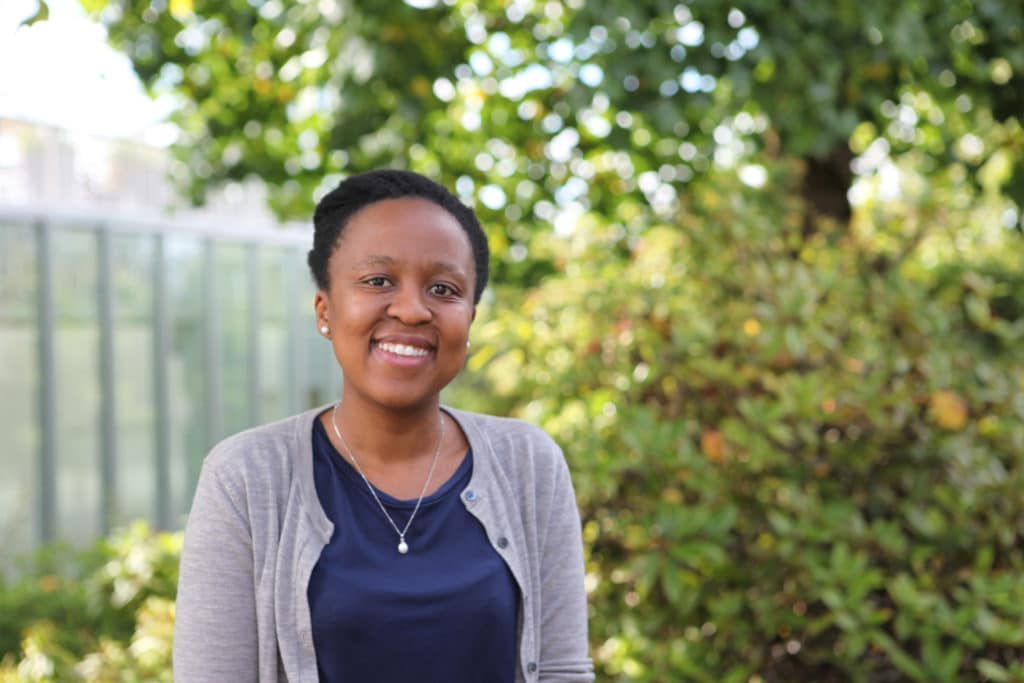
“COVID-19 offered a unique opportunity to test drive our models to try to understand how COVID-19 changes the coagulation system in the body,” said Malebogo Ngoepe, associate professor in the Department of Mechanical Engineering at the University of Cape Town and STIAS Iso Lomso fellow. Ngoepe was presenting her work at the first STIAS public webinar of the current semester.

‘We know that the spike proteins on COVID-19 allow it to enter the body by attaching to host cells via the angiotensin-converting enzyme 2 (ACE2) receptor, of which there is an abundance on endothelial cells. COVID-19 causes disruption of the endothelium and, with damage, clotting can occur. We wanted to understand what happens when COVID-19 causes clotting.”
Compared to clot formation in healthy plasma they found that in COVID-19, due to changes in blood viscosity, significant clots formed in a very short time period – 90 seconds – and that these reduced blood flow.
“Also when we washed out the channels it was much harder to remove the COVID-19 clots even from the plastic medium. So dissolving them is clearly a huge issue which affects which drugs might be effective.”
“Next we want to look at the effect of the different COVID-19 variants on clotting.”
Looking at blood flow and clotting in COVID-19 is a natural progression of Ngoepe’s modelling work on aneurysms, thrombosis and congenital heart conditions.
So what has mechanical engineering to do with blood?
“At a basic level the heart is a pump and the blood vessels are pipes,” said Ngoepe.
She explained therefore that computational and experimental flow techniques, commonly used to study fluid flows in more traditional engineering contexts, can be successfully translated to medical and biological applications. Computational fluid dynamics is an important technique for the development of models enabling insight into blood clotting in various diseases.
“Thrombosis, or blood-clot formation, is an important feature of many vascular diseases,” she said. “This process is influenced by many variables, including biochemical reactions and blood flow. Many of the breakthroughs in our understanding of blood clots have been led by the biochemistry and physiology communities. The addition of haemodynamics, or blood flow, is a relatively recent development which contributes to our understanding of blood-clot formation in disease.”
Virchow’s Triad, first described by Rudolph Virchow in the mid-1800s, showed that clotting is a balance between blood constitution, the vessel wall and flow. This balance is carefully maintained and only deviates under conditions of disease.
“Under normal conditions an intact endothelium separates the layers in the blood vessel from the flowing blood and there is no spontaneous clotting,” explained Ngoepe. “With injury, there is disruption to the endothelial sub layers, they become exposed and there may be a gap in the wall.”
Aneurysms, balloon-like expansions of blood vessel walls caused by the weakening of the layers, may form. These are most commonly found on blood vessels of the brain and on the aorta, and are at risk of rupture with subsequent morbidity or mortality. They are also linked with clot formation. Clots that grow in aneurysms can either assist by sealing off the aneurysm, can exacerbate the situation by speeding up the time to rupture, or can break away causing problems elsewhere in the body.
Current treatments for aneurysms involve stents, coils and flow diverters. These reduce the flow of blood to the aneurysm sack and reduce the possibility of rupture. However, they can also cause clotting and, in some cases, speed up the time to rupture.
Ngoepe pointed out that for most people aneurysms don’t actually rupture and “they just live with it”. However, when they are picked up – often during other medical investigations – the doctor has to make the difficult decision to treat, which may be unnecessary and could worsen the situation, or to leave it alone which can prove fatal if it ruptures.
“Doctors don’t treat every aneurysm – operating exposes patients to unnecessary risk if it never ruptures.”
“Aneurysms are different in every patient – if you look at the aneurysm geometry from MRI scans you see this. Clots also differ substantially so we need an indication of what type of clot will form.”
Ngoepe and her colleagues have therefore been working on a computational model which uses fluid flow and biochemical modelling to predict clot growth in aneurysms. The computational aspects include the ability to reconstruct a 3D-printed version of the blood vessels and aneurysm geometry, as well as a biochemical model which describes the patient’s unique clotting process based on different concentrations of proteins in the blood and other variables.
“The biochemical aspect allows us to understand the reactions between the different chemicals in the blood. Models need to account for both the fluid flow and biochemical components as well as allowing comparison between treatment vs. no treatment.”
But how do we know that a computer model really replicates reality?
“We must verify,” said Ngoepe. In order to ensure that the computer model outputs are in an understandable format and that the model replicates clinical reality, Ngoepe and researchers from UNISA have also developed an experimental model that physically maps the fluid-flow path through the vessel and aneurysm.
Using particle image velocimetry, a non-intrusive laser optical measurement techniqueresults to the computer model confirming the reality of the model.
“We also figured out how to grow clots using thrombin and fibrinogen and tracked the clot growth,” said Ngoepe. “We are now looking at tracking the flow field and clotting simultaneously.”
In addition to brain aneurysms these models are used to understand abdominal aortic aneurysms which affect vessels directly flowing from the heart and are affected by the heart’s pumping action which causes a different clotting profile as well as deep vein thrombosis which form in veins and therefore involve deoxygenised blood and also the challenge of opening and closing valves.
“The models are also proving useful in cases of children with heart disease – where we look at the fluid field and try to predict what will happen if you operate. The challenge with children is that they will grow affecting any intervention and you also want to operate only once with the best outcomes. So we run simulations to try to help the doctor’s decision making.”
But Ngoepe was quick to emphasise that models never replace experience.
“Such models are value adding,” she said. “They are tools and techniques that give some insights but more crucial is the doctor’s experience and clinical knowledge.”
“Getting models into the clinical space is a long road,” she added. “You first have to develop a common language with the doctors.”
“It’s also about balancing accuracy against time. This provides a tool for clinicians to get insight into the clotting procedure in quick time. There is a need to use this as a starting point to understand longer time periods. Clots in aneurysms, for example, usually develop over a 12 – 18 month period. ”
It takes a village
Ngoepe’s exciting work over the last number of years is now being put to use in the huge challenge currently facing the world – the COVID-19 pandemic. She is hoping the models will allow greater insight into the process of clotting initiated by COVID-19, the growth and activity of the clots, why some people with COVID-19 are more susceptible to clotting, and what drugs will be effective at dissolving resistant COVID-19 clots.
“People generally clot too much or too little. Some people are clearly more at risk for developing COVID-related clots but there are many factors and complicated interactions involved.”
“We hope these models could give a platform to test quite quickly and easily at an experimental level what might dissolve a clot. Many of these drugs are currently being tested in large clinical trials.”
But it’s all about looking at the problem from every angle and Ngoepe’s work clearly highlights the huge importance of an interdisciplinary approach to the global challenges we face.
Michelle Galloway: Part-time media officer at STIAS
Photograph: supplied
